

11 December 2024: The Use of AI to Improve Access to High-Quality Radiation Therapy Treatment Planning in Low- and Middle-Income Countries
11 December 2024 at 12 pm GMT; Duration 1 hour
Moderator: M. Mahesh, MS, PhD. Chair of IOMP Science Committee
Speakers:
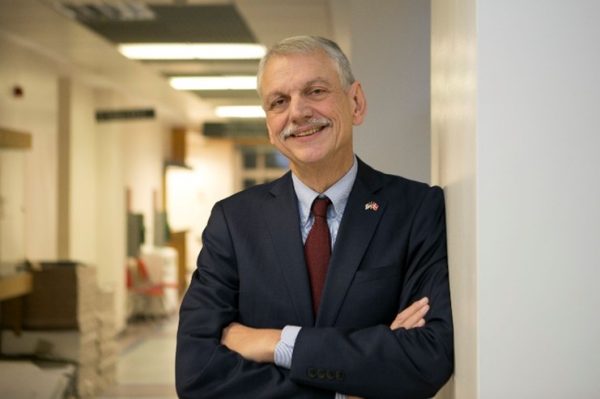
Laurence Court, PhD, University of Texas MD Anderson Cancer Center, USA
Barbara Marquez, University of Texas MD Anderson Cancer Center, USA
Christoph Trauernicht, PhD, Tygerberg Hospital / Stellenbosch University, South Africa
Abstract:
Advances in artificial intelligence (AI) are going to affect all aspects of radiation therapy, including contouring, treatment planning, and quality assurance. This presentation will describe our efforts to develop and expand these tools specifically for clinics in low- and middle-income countries. We will describe the expected quality (clinical acceptability, efficiency gains) of these tools, as well as possible risks in deployment and how to mitigate them. Finally, we will describe lessons learned in clinical deployment.
Learning Objectives:
- To be able to describe how AI is likely to improve access to high-quality radiotherapy planning across the world.
- To understand risks when implementing AI into clinical practice, and possible ways to reduce them.
- To understand challenges in implementing these tools into clinical practice in LMICs.
Speakers’ CV:
Laurence leads a team who are focused on the development and clinical deployment of AI-based tools to support radiotherapy teams in low- and middle-income countries. Their flagship product is the Radiation Planning Assistant, which received FDA 510(k) clearance in 2023, and is currently starting clinical use in South Africa.
Christoph Trauernicht is the Head of the Medical Physics Division at Tygerberg Hospital, Cape Town, South Africa, and a Senior Lecturer at Stellenbosch University. He leads a team of five medical physicists, two radiographers and a mould room, together with a couple of medical physics clinical training posts.
28 October 2024: Why Radiation Oncology Clinical Trials need Medical Physicists
28 October 2024 at 12 pm GMT; Duration 1 hour
Moderators:
Wayne Beckham, PhD, FCCPM, FCOMP, British Columbia Cancer, Victoria, Canada
Kwan Hoong Ng, PhD, DABMP, FIOMP, FIUPESM, Universiti Malaya, Kuala Lumpur, Malaysia
Speakers:
Søren M. Bentzen, Ph.D., D.M.Sc., FBIR, FASTRO, University of Maryland, Baltimore, USA
Tomas Kron, OAM, Ph.D., FCCPM, FACPSEM, FIOMP, Peter MacCallum Cancer Centre, Melbourne, Australia
Abstract:
Randomized controlled trials (RCT) remain the gold standard for establishing causality between an intervention and a clinical outcome. Leading into an RCT is a hierarchy of early phase clinical trials. While trial data are typically generated under highly controlled conditions in selected patient populations, the data are prospectively acquired and of high quality which is important to establish a proof-of-concept. Radiation Oncology has a proud history of conducting clinical trials and medical physicists play an essential role in reducing the patient-to-patient variability in exposure that is not attributable to the intervention itself. Some key roles for medical physicists in trials are:
- Defining the technical details of the radiotherapy approach used in the trial
- Independent credentialling of centres participating in the trial
- Developing a quality assurance program to ensure collection of high quality data
- Supporting (automated) data collection, curation, and evaluation
- Developing trial questions that could help to identify the role of techniques or technologies in improving patient outcomes.
- Many medical physicists are not aware of the importance of trials and the opportunities they provide to create relevant evidence and engage with clinicians. The presentation aims to provide background, examples, and motivation for this.
Learning Objectives:
- Be aware of different approaches to clinical trial
- Understand the importance of clinical trial for generation of evidence in radiation oncology
- Appreciate the multiple roles medical physicists can have in clinical trial
- Explore where this can result in more than a supporting role
Speakers’ CV:
Søren M. Bentzen, Ph.D., D.M.Sc., FBIR, FASTRO
Søren is Professor and Director of the Division of Biostatistics and Bioinformatics, and Professor of Radiation Oncology at University of Maryland School of Medicine. He was born and educated in Denmark and started out as a hospital physicist in radiation oncology. He earned a PhD in medicine (quantitative medical imaging) and later a Doctor of Medical Science in clinical radiobiology. Søren spent his research career at the interface between radiation biology, medical physics, biostatistics and evidence-based radiation oncology, working previously at University of Aarhus, Denmark; MD Anderson Cancer Center; the Gray Laboratory, University College London; University of Wisconsin – Madison. Søren has published 550 papers and book chapters and presented 400 invited lectures. His papers have been cited 50,000 times and his h-index is 112 (Google Scholar). He received 34 awards and honours and is one of only 3 individuals awarded both the ASTRO and ESTRO Gold Medals.
Tomas Kron, OAM, Ph.D., FCCPM, FACPSEM, FIOMP
Tomas Kron was born and educated in Germany. After his PhD he migrated to Australia in 1989 where he commenced his career in radiotherapy physics. He now is Director of Physical Sciences at Peter MacCallum Cancer Centre in Melbourne leading a team of some 50 engineers and physicists. Tomas has an interest in education of medical physicists, dosimetry of ionising radiation, image guidance and clinical trials, which is demonstrated by more than 100 invited conference presentations and 350 papers in refereed journals. He has been principal investigator in several clinical trials aiming at demonstrating feasibility of various technical approaches to radiotherapy treatment and has received an Order of Australia Medal for services to science and medicine.
3 July 2024: Advancing Radiation Safety in Medicine: Insights from IOMP and UNSCEAR on Ionizing Radiation Exposure
3 July 2024 at 12 pm GMT; Duration 1 hour
1 CPD/CME point is applied to this event.
Organizer: John Damilakis, President IOMP
Moderator: John Damilakis, President IOMP
Title 1: Setting the stage: A short introduction to IOMP’s role in enhancing radiation safety in medical practices
Speaker: John Damilakis, MSc, PhD, FIOMP, FIUPESM
President, IOMP
John Damilakis is a professor and director of the Department of Medical Physics, School of Medicine, University of Crete and director of the Department of Medical Physics of the University Hospital of Heraklion, Crete, Greece. He holds the position of President within the ‘International Organization for Medical Physics’ (IOMP) and has previously held leadership roles, including President, in organizations such as the ‘European Federation of Organizations for Medical Physics’ (EFOMP), the ‘European Alliance for Medical Radiation Protection Research’ (EURAMED), and the ‘Hellenic Association of Medical Physics’.
Professor Damilakis also contributes to international initiatives, serving as a member of the International Commission on Radiological Protection (ICRP) Committee 3, Chair of the International Union of Pure and Applied Physics (IUPAP) AC4 and participating in the steering committee of the ‘EuroSafe Imaging Campaign’ organized by the European Society of Radiology. He has shared his expertise as a Visiting Professor, delivering lectures on medical dosimetry and medical radiation protection at Boston University in the United States.
His body of research work encompasses a wide range of research areas, including medical dosimetry, medical radiation protection, and the application of artificial intelligence in medical imaging. He has played significant roles as an editor, author, or co-author in several influential books within the field. Professor Damilakis has published 260 research articles listed on PubMed, accompanied by 9728 citations, and an h-index of 53 as documented on Google Scholar (May, 2024). His contributions to the field have garnered him numerous awards and recognitions for his scientific achievements.
Title 2: Insights and future directions in medical radiation exposure: A detailed analysis from UNSCEAR’s 2020/2021 Report
Speaker: Peter Thomas

Dr Peter Thomas is the director of the Medical Imaging section at the Australian Radiation Protection and Nuclear Safety Agency (ARPANSA). The section runs the Australian National Diagnostic Reference Level Service, provides educational material and advice, contributes to codes and safety guides, and promotes optimal use of medical radiation. Dr Thomas served as the chair of the UNSCEAR Expert Group on Medical Exposures from 2019 to 2020 and was a lead writer for the 2020/2021 UNSCEAR report summarising the global level of ionizing radiation exposures in medicine. Dr Thomas was a member of the Australian delegation to UNSCEAR in 2005 and from 2019 to 2024. Dr Thomas has a PhD in Chemistry from Monash University and has worked at ARPANSA since 2000.
Abstract:
This webinar focuses on critical developments and findings in the field of medical exposure to ionizing radiation. The webinar will begin with a brief introduction by IOMP, presenting IOMP’s role in enhancing radiation safety across medical practices globally, key areas of focus in ionizing radiation medical exposure, and the importance of collecting data related to frequency, distribution, and trends of radiation usage in medical diagnostics and treatment. Following this, a detailed presentation by UNSCEAR will focus on the comprehensive “Evaluation of Medical Exposure to Ionizing Radiation” report. The webinar aims to equip participants with insights into the global state of medical radiation practices, advancements in the field, and upcoming evaluations by UNSCEAR.
Learning Objectives
For IOMP’s presentation:
- To understand the role of IOMP in promoting radiation safety and effective practices in medical imaging.
- To identify key areas and recent advancements in radiation protection in medical settings.
- To discuss the importance of global collaboration in improving patient and staff safety.
For UNSCEAR’s presentation:
- To present key findings from the UNSCEAR 2020/2021 Report regarding medical exposure to ionizing radiation.
- To discuss trends in radiation exposures in medicine noted by UNSCEAR and any research needs
- To present UNSCEAR’s plans for a future evaluation.
20 June 2024: (1) Advancements in Imaging for Radiation Therapy and (2) Patient Specific QA in IMRT
20 June 2024 at 12 pm GMT; Duration 1 hour
Organizer: Eva Bezak
Moderator: Ibrahim Duhaini
Title 1: Advancements in Imaging for Radiation Therapy
Speaker: Hassaan Alkhatib, PhD, FAAPM, DABR, AABMP
Chief Physicist at South Carolina Oncology Associates, Columbia, South Carolina, USA
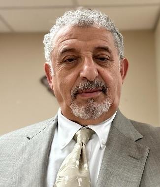
Dr. Hassaan Alkhatib is the Chief Physicist at South Carolina Oncology Associates, Columbia, South Carolina. He earned his doctorate degree from the University of North Dakota and completed his Medical Physics Residency at the University of Minnesota. Subsequent to his doctorate degree in Health Physics in 1996, Hassaan has accumulated considerable academic and professional experience. His medical physics educational activities include his educating and training of medical physics students, residents, dosimetrists, and radiation therapists for about 20 years. He continues his teaching and training activities at Richland Memorial Hospital, Columbia, South Carolina, where he has been Chief of Medical Physics since 2005 and Associate Professor at the University of South Carolina since 2010. He obtained board certification from American Board of Medical Physics (AABMP) and American Board of Radiology (ABR).
Dr. AlKhatib has contributed significantly through teaching, clinical service, research, administration, and participation in the AAPM professional and educational activities. He is also Physics Advisory Board Member in Oncology Data Systems Inc. He is expertise as a scientist, a teacher and author/co-author of many scientific publications..
Abstract:
This seminar explores the latest developments in imaging technologies for optimizing radiation therapy treatments. From traditional methods to cutting-edge innovations, participants will gain insights into how imaging enhances precision, accuracy, and patient outcomes in radiation oncology. The seminar will cover key concepts, practical applications, and future directions in the field.
Learning Objectives:
Knowledge:
- Understand the fundamentals of imaging modalities used in radiation therapy, including X-ray, CT, MRI, PET, and ultrasound.
- Explore the principles behind image acquisition, reconstruction, and processing techniques specific to radiation therapy planning and delivery.
- Gain insights into emerging imaging technologies such as image-guided radiation therapy (IGRT), adaptive radiotherapy, and functional imaging for treatment response assessment.
Skills:
- Develop proficiency in utilizing imaging data for target delineation, treatment planning, and dose optimization in radiation therapy.
- Acquire skills in image registration, fusion, and co-registration methods to integrate multiple imaging modalities for comprehensive treatment planning.
- Learn techniques for image-guided setup verification, motion management, and real-time monitoring during radiation treatment delivery.
Competences:
- Enhance critical thinking and problem-solving abilities in assessing imaging data quality, interpreting anatomical and functional information, and adapting treatment strategies accordingly.
- Foster effective communication and collaboration between radiation oncologists, medical physicists, radiation therapists, and imaging specialists to optimize patient care and treatment outcomes.
- Cultivate a mindset of continuous learning and staying updated with advancements in imaging technology, radiation therapy techniques, and clinical evidence through research, education, and professional development initiatives.
Title 2: Patient Specific QA in IMRT
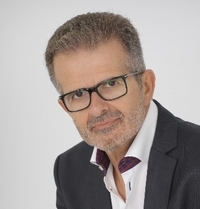
Speaker: Dr. Sudesh Deshpande, Ph.D.
Chief Medical Physicist, P.D. Hinduja National Hospital and Research Center, Mumbai
Education:
• B.Sc. (1993), M.Sc. (1996), Ph.D. (2016) – Amravati University, Maharashtra
• D.R.P. (1997) – Mumbai University, Maharashtra
Total experience 27 years in the field of Medical Physics
Appointments:
• Jr. Medical Physicist, Cancer Hospital and Research Institute, Gwalior (1997-1999)
• Jr. Medical Physicist, P.D. Hinduja National Hospital, Mumbai (2000-2001)
• Medical Physicist, Kailash Cancer Hospital, Goraj (2001-2002)
• Medical Physicist, Tata Memorial Hospital, Mumbai (2003-2006)
• Chief Medical Physicist, P.D. Hinduja National Hospital, Mumbai (2006-present)
Areas of Interest:
• Clinical: Advancing external beam therapy, implementing 3D treatment planning, IMRT, SBRT.
• Research: Imaging dose optimization.
Publications: 25
Professional Services:
• Executive Committee, AMPI (2011-2013, 2024-2027)
• Member, AERB Safety Review Committee (2017-2023), Standing committee for Document development
• Member of Education and training committee of AFOMP (2023-2025)
• Reviewer for multiple scientific journals
Academic Activities:
• Guest lecturer for Dip R P BARC students
• Invited speaker at national and international conferences
• Course Coordinator for VMAT-IMRT and TrueBeam physics courses at Hinduja Hospital
• Member of task group for standardisation of QA procedure for advanced radiotherapy
Specialized Work:
• Commissioning of various accelerators and treatment planning systems
• Implementation of advanced radiotherapy techniques and technologies
Affiliations:
• Life member, Association of Medical Physicist of India
• Life member, Indian Brachytherapy Society
Abstract:
Patient-specific quality assurance (PSQA) in Intensity-Modulated Radiation Therapy (IMRT) is important step to ensure treatment accuracy and patient safety. The importance of patient-specific QA lies in its ability to detect and correct errors specific to the treatment plan, machine performance thus enhancing the precision and effectiveness of IMRT. PSQA contains point dose measurement and two-dimensional (2D) dose distribution measurements for point dose measurements, the choice of detector is crucial. Ionization chambers are often preferred due to their high accuracy and stability, making them ideal for absolute dose verification. For 2D dose distribution measurements, arrays of ionization chambers, diodes, or electronic portal imaging devices (EPIDs) are commonly used. These 2D detectors provide comprehensive spatial resolution, enabling the detection of dose discrepancies across the entire treatment field.
The AAPM’s Task Group 218 (TG-218) provides recommendations to standardize and improve patient-specific QA procedures. It emphasizes the use of gamma index analysis with criteria of 3% dose difference and 2 mm distance-to-agreement (3%/2 mm) for evaluating dose distributions. TG-218 also suggests thresholds for passing rates to ensure high-quality treatments.
Tips and tricks for effective QA include regular calibration of detectors, verification of treatment plan calculations, and cross-validation with multiple QA tools. Additionally, integrating software solutions for automated analysis can enhance the efficiency and reliability of QA processes. Adopting these strategies ensures robust patient-specific QA, ultimately contributing to the success of IMRT treatments.
Learning Objectives:
- To understand importance of Patient specific QA in IMRT
- Choice of detectors for point dose and 2D dose distribution measurements
- Use of AAPM TG 218 for PSQA analysis
- Tips and Tricks for PSQA
- New trends in PSQA in IMRT
29 May 2024: Quality Assurance in Radiation Therapy
29 May 2024 at 12 pm GMT; Duration 1 hour
1 CPD/CME point is applied to this event.
Organizer and Moderator: Eva Bezak, Vice President IOMP
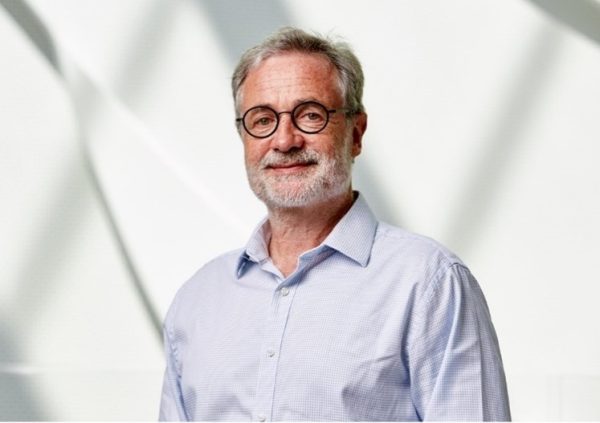
Speaker: Alexandru Dasu
The Skandion Clinic, Uppsala, Sweden.
Department of Immunology, Genetics and Pathology, Uppsala University, Uppsala, Sweden.
Alexandru Dasu is the Chief Medical Physicist at the Skandion Clinic, the national proton therapy centre in Uppsala, Sweden and academically affiliated to Uppsala University. Alexandru Dasu studied Medical Physics at Umeå University, Sweden, where he also received a Ph.D. degree in 2001. He became a certified medical physicist in 2003 and throughout his career combined clinical activities with research and education. His main research interests include proton therapy, the evaluation of the risk for stochastic effects following radiation therapy, the modelling of the tumour response to radiation therapy, radiation dosimetry and quality assurance.
Contact details:
Alexandru Dasu
The Skandion Clinic
Uppsala, Sweden
E-mail: alexandru.dasu@skandion.se
Abstract:
Radiation therapy is a multi-disciplinary, multi-step treatment using complex equipment and techniques to deliver ionizing radiation generally for cancer patients. Each step in the process can be subject to constraints and variation that can affect the fine balance between delivering enough dose to the target while keeping the doses to the normal tissues at acceptable levels. Indeed, variations above 5% of the dose can lead to significant changes in local control or complication rates, this worsening the effectiveness of the treatment. A rigorous quality assurance system is therefore needed to ensure that the accurate doses have been delivered in the defined volume within the scheduled time and with limited dose to surrounding normal tissues.
This presentation will review the rationale for quality assurance in radiation therapy, the particular requirements of radiation therapy versus radiation protection, the steps to build a quality assurance as well as examples of the steps allowing a continuous improvement of quality in radiation therapy.
Learning Objectives
After the presentation, the attendees should be able to understand the rationale for quality assurance in radiation therapy and the methodology for setting up a quality assurance program, as well as the relationship between quality assurance and quality controls.
8 Mar 2024: Celebrating International Women’s Day with Early Career Medical Physicists
8 March 2024 at 12 pm GMT; Duration 1 hour
1 CPD/CME point is applied to this event.
Organizer: Loredana Marcu, Eva Bezak & Kathleen Hintenlang
Moderator: Loredana Marcu
Title 1: In vitro development of MUC1-CE targeted alpha therapy for pancreatic ductal adenocarcinoma
Speaker: Ashleigh Hull
Allied Health and Human Performance Academic Unit, University of South Australia, Adelaide, SA, Australia.
Department of Nuclear Medicine, PET and Bone Densitometry, SA Medical Imaging, Royal Adelaide Hospital, Adelaide, SA, Australia.
Ashleigh Hull is a lecturer in Nuclear Medicine and PhD candidate (under submission) at the University of South Australia. Ashleigh’s research focuses on the development and characterisation of novel radioimmunoconjugates for the diagnosis and treatment of cancer. Her PhD project has led to the development of two novel therapeutic radioimmunoconjugates capable of treating pancreatic cancer at an in vitro level. In addition to her academic and research roles, Ashleigh also maintains her clinical practice as a registered nuclear medicine technologist at SA Medical Imaging.
Abstract:
Pancreatic ductal adenocarcinoma (PDAC) is a highly aggressive malignancy and a leading cause of cancer-related death worldwide. Development of targeted therapies, such as a targeted alpha therapy (TAT), may improve outcomes of patients diagnosed with PDAC. In this study, a novel alpha-emitting radioimmunoconjugate, 225Ac-DOTA-C595, was developed to target PDAC by binding to cancer-specific mucin 1 epitopes (MUC1-CE). MUC1-CE is known to be overexpressed on over 90% of PDAC tissues yet has minimal expression on normal and benign tissues, offering an ideal expression profile for TAT purposes. A series of in vitro assays were performed to evaluate cell binding, internalisation and cytotoxicity of 225Ac-DOTA-C595 and establish the feasibility of using 225Ac-DOTA-C595 as a radioimmunoconjugate for the treatment of PDAC.
Title 2: Exploring Ultra-high Dose Rate Radiation and Growing as an Educator in Medical Physicist
Speaker: Ashley Cetnar, PhD, Assistant Professor Radiation Oncology
Ashley Cetnar is an Assistant Professor in the Department of Radiation Oncology at The Ohio State University. She is trained as both a clinical medical physicist and expert in teaching and learning. Within the clinic, she strives to provide excellent patient care in her role as a medical physicist by solving problems, applying science and technology, upholding quality and safety, and promoting innovation for new ways of treating patients. As an educator, she is passionate about helping others learn and grow as both individuals and within a team.
Abstract:
In this presentation, Dr. Cetnar will share her journey of involvement of research in ultra-high dose rate radiation therapy and journey professionally growing as an educator. From a research perspective, The Ohio State University currently has two ultra-high dose rate generating devices that can be used to deliver electrons with the next expected expansion to protons soon with the opening the new of proton therapy facility. This will include background information about FLASH, commissioning of new technology, exploration of dosimetry methods, and biological experiments. Dr. Cetnar has always been passionate about education and will share her pathway to completing a PhD in Education, educational programs, and medical physics education research.
Title 3: Making progress only needs to get started
Speaker: Leticia Irazola Rosales, Medical Physics at Centro de Investigaciones Biomédicas de la Rioja
Leticia Irazola is a Medical Physics from Spain. She made her studies in Physics at the University of Zaragoza (Spain) and then dedcated to the Medical Physics field with her MSc in Medical Physics at the University of Rennes I and the Centre Eugène Marquis (France), PhD thesis at the University of Sevilla (Spain) and PostDoc at the Pontificia Universidad Católica de Chile, Santiago de Chile (Chile).
Then she moved into the clinical field by performing the Medical Physics residency at the Clinica Universidad de Navarra (Spain). Based in La Rioja (Spain), she works now as a medical physicist combining her job as a Medical Physicist with university teaching and research at the Centro de Investigaciones Biomédicas de La Rioja.
She is currently secretary of the Communications and Publications Committee, Secretary of the Spanish Early Career group of the SEFM and Chair of the Early Career SIG of the EFOMP.
Abstract:
I have never in my youthfulness imagined that I would became the Secretary or Chair of any scientific group.
When I was studying my University Degree I just thought which would my future become by studing such a “theoretical” career as Physics. During my 2nd year at University I assisted to a talk related to the Medical Physics profession and I realized then that it was the path I wanted to follow, I felt it was just puting my humble knowledge of physics to the service of public health.
Since then I can almost say that all my life has been by chance and made by choice. What I have clear at this point is that it is not only a matter of being the best at doing something but loving what you do and being passionate about it. This is the way best things come to you and dreams become reality.
24 Jan 2024: From Pixels to Patients: The Influence of Gaming and Smartphone Developments on Radiation Oncology
24 January 2024 at 12 pm GMT; Duration 1 hour
1 CPD/CME point is applied to this event.
Organizer: Eva Bezak
Moderator: Eva Bezak
Title: From Pixels to Patients: The Influence of Gaming and Smartphone Developments on Radiation Oncology
Speaker: Dr Michael Douglass, Royal Adelaide Hospital, Adelaide, Australia
Dr. Michael Douglass is a principal medical physicist at the Royal Adelaide Hospital where he oversees treatment planning systems and planning support services. He also works part-time at the Australian Bragg Centre for Proton Therapy and Research where he contributes to proton therapy comparative planning and research. In addition, Dr. Douglass holds an academic role at the University of Adelaide, supervising Ph.D. and master’s student research. His expertise spans across a multitude of research areas, including proton therapy, Monte Carlo simulations, machine learning and 3D printing in radiation oncology, which is reflected in his 40+ peer-reviewed publications. Dr. Douglass’s notable contributions to the field have earned him multiple awards such as the 2021 Simpson Prize for Cancer Research and the 2021 ACPSEM Boyce Worthley Young Achiever Award.
Abstract:
In this presentation, we will explore how advancements in various industries, including the movie, video game, and smartphone industries, have contributed to the progress of radiation oncology. We will discuss how developments such as visual effects in the movie industry have paved the way for the creation of synthetic training data for machine learning models in radiation oncology. Additionally, we will examine how the smartphone industry has enabled new methods of 3D scanning patients for treatments like TBI, TSET, Brachytherapy, and 3D printed bolus, thanks to technologies like Gaussian platting, NERF, LiDAR, and iPhone capabilities. The integration of augmented reality in the video game industry has revolutionized the visualization of medical imaging data, providing volumetric views and assisting in breath hold coaching. We will also explore the impact of GPU technology on deep learning segmentation, auto planning, and accelerated dose calculations, as well as the potential of large language models like Llama 2 and GPT in education and automated patient record transcription. Through these examples, we will highlight the interconnectedness of these industries with radiation oncology and the ongoing advancements they bring to the field.
Learning Objectives:
- Gain an understanding of the advancements in optical scanning technologies, including LiDAR, photogrammetry, and NERF, and their applications in radiation oncology.
- Understand how consumer technologies, such as smartphones and gaming, have contributed to the progress of radiation oncology and medicine as a whole.
- Learn about the benefits and potential applications of large language models, like Llama 2 and GPT, in radiation oncology, including education and automated patient record transcription.
- Examine the integration of visual effects in the movie industry and its influence on the creation of synthetic training data for machine learning models in radiation oncology.
- Understand the impact of augmented reality in the video game industry on the visualization of medical imaging data and its role in assisting in breath hold coaching.